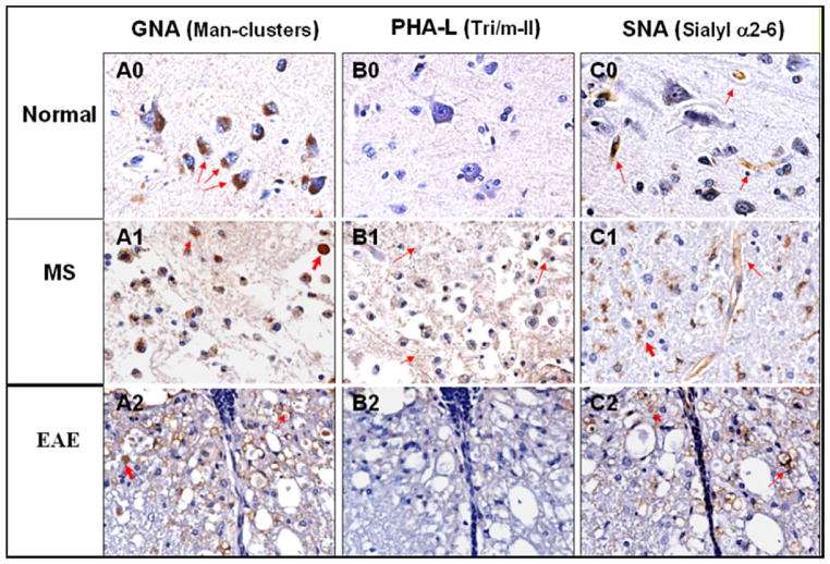
Fig. 5.
Lectin GNA, PHA-L and SNA histochemistry of human brain tissues, normal (0) and MS (1), and mouse spinal cord EAE lesions (2). CNS tissues were stained with biotinylated lectins and developed with Streptavidin-HRP conjugate and DAB substrate. Lectins were pre-titrated and applied at 10 μg/ml for tissue staining. Column A, B, and C were stained with lectin GNA, PHA-L and SNA respectively. Normal: A0, B0, and C0; MS: A1, B1, and C1; EAE: A2, B2, and C2. The carbohydrates recognized by the lectins are indicated for each column.

