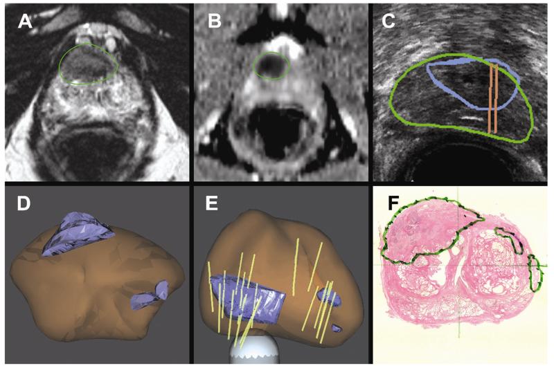
Fig. 3.
A 65-year-old man with PSA 8.5 on active surveillance after initial biopsy showed 1 mm of Gleason 3 + 3 cancer in 1 core. (A and B) Anterior lesion of highest suspicion identified on mpMRI. (C) Real-time US targeting of the corresponding lesion. (D and E) 3D models demonstrate the target (blue), prostate (brown), and biopsy cores (tan cylinders). Note that in panel E the 3D model is rotated, making the anterior tumor appear to be posterior. MR-US fusion confirmatory biopsy revealed Gleason 4 + 3 cancer. (F) Radical prostatectomy pathology confirmed a 2.3 cm Gleason 4 + 4 cancer centered in the right, anterior prostate. (Color version of figure is available online.)

