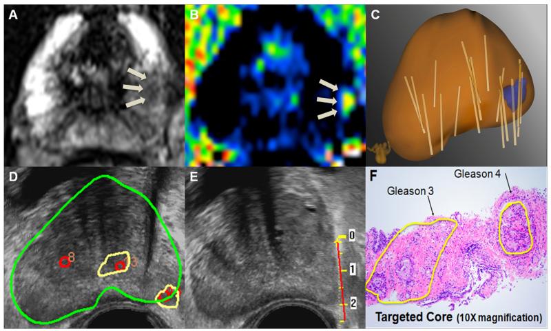
Fig. 5.
Illustrative case of a 66-year-old man with a PSA of 9 ng/mL. MRI demonstrates 2 image grade 3 targets. (A) T2-weighted MRI shows a left-sided lesion with moderately reduced signal. (B) Diffusion-weighted imaging confirms moderately restricted diffusion in the area of interest. (C) Fusion of US and MRI generates a 3D model. Individual biopsy cores (tan cylinders) are mapped on the model. (D) Targets are superimposed on real-time US to enable targeted biopsy. (E) Real-time tracking of biopsy cores ensures accurate biopsy of targeted lesions. (F) In this case, diagnostic Artemis fusion biopsy showed one core of Gleason 3 + 3 (1 mm) on systematic biopsy, thereby fulfilling Epstein criteria. However, targeted biopsy showed a focus of Gleason 3 + 4. Based on systematic biopsy alone, this patient would be considered an excellent candidate for active surveillance. Inclusion of targeted biopsy and application of current risk models would result in upstaging to intermediate risk and perhaps definitive treatment. Further study will be required to develop new risk assignment criteria based on targeted biopsy and to determine if men with small, Gleason 3 + 4 tumors on targeted biopsy are suitable for active surveillance. (Color version of figure is available online.)

