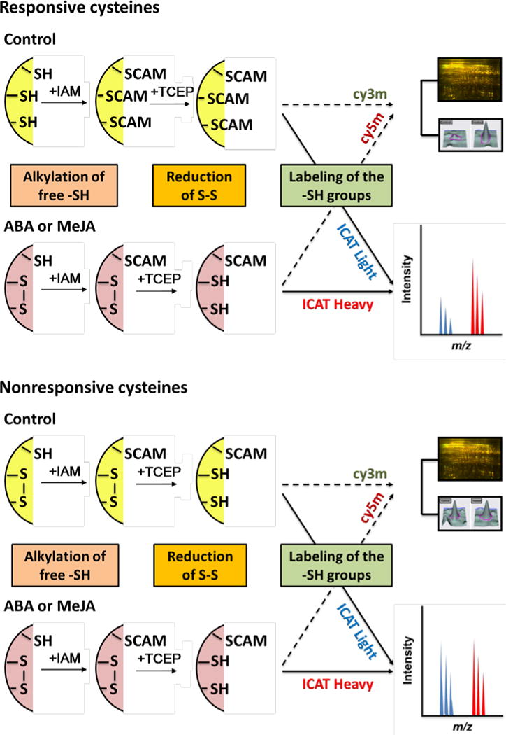
Figure 1.

Simplified diagram showing complementary approaches of saturation DIGE and ICAT used to identify redox sensitive proteins. Proteins from control and hormone-treated guard cells were first alkylated to block remaining free -SH groups, then the cysteines oxidized were reduced and labeled with Cy dyes or ICAT reagents, followed by DIGE and LC-MS/MS. IAM, iodoacetamide; CAM, carbamidomethylation; TCEP, tris(2-carboxyethyl)phosphine.
