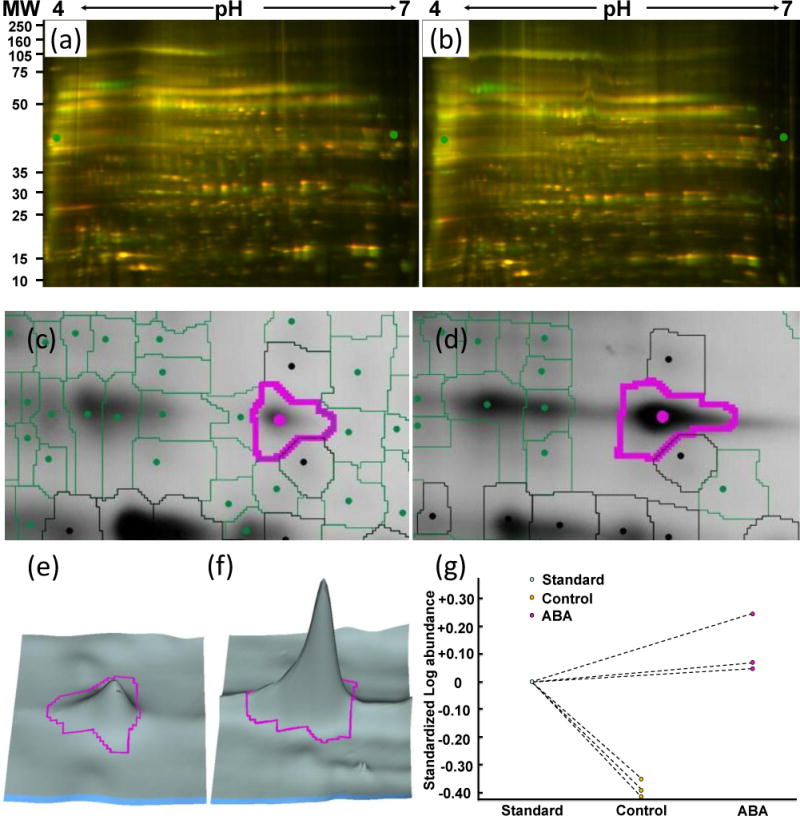
Figure 2.

Example of redox protein identification using the DIGE approach. (a) DIGE image of control guard cell proteins. (b) DIGE image of ABA treated guard cell proteins. (c) A protein spot from control sample. (d) The same protein spot from ABA-treated sample showing its redox regulation. (e) 3D view of (c). (f) 3D view of (d). (g) Quantitative changes of the spot across replicate samples. The protein spot was identified as myrosinase Myr2 (gi414103).
