
Figure 3.
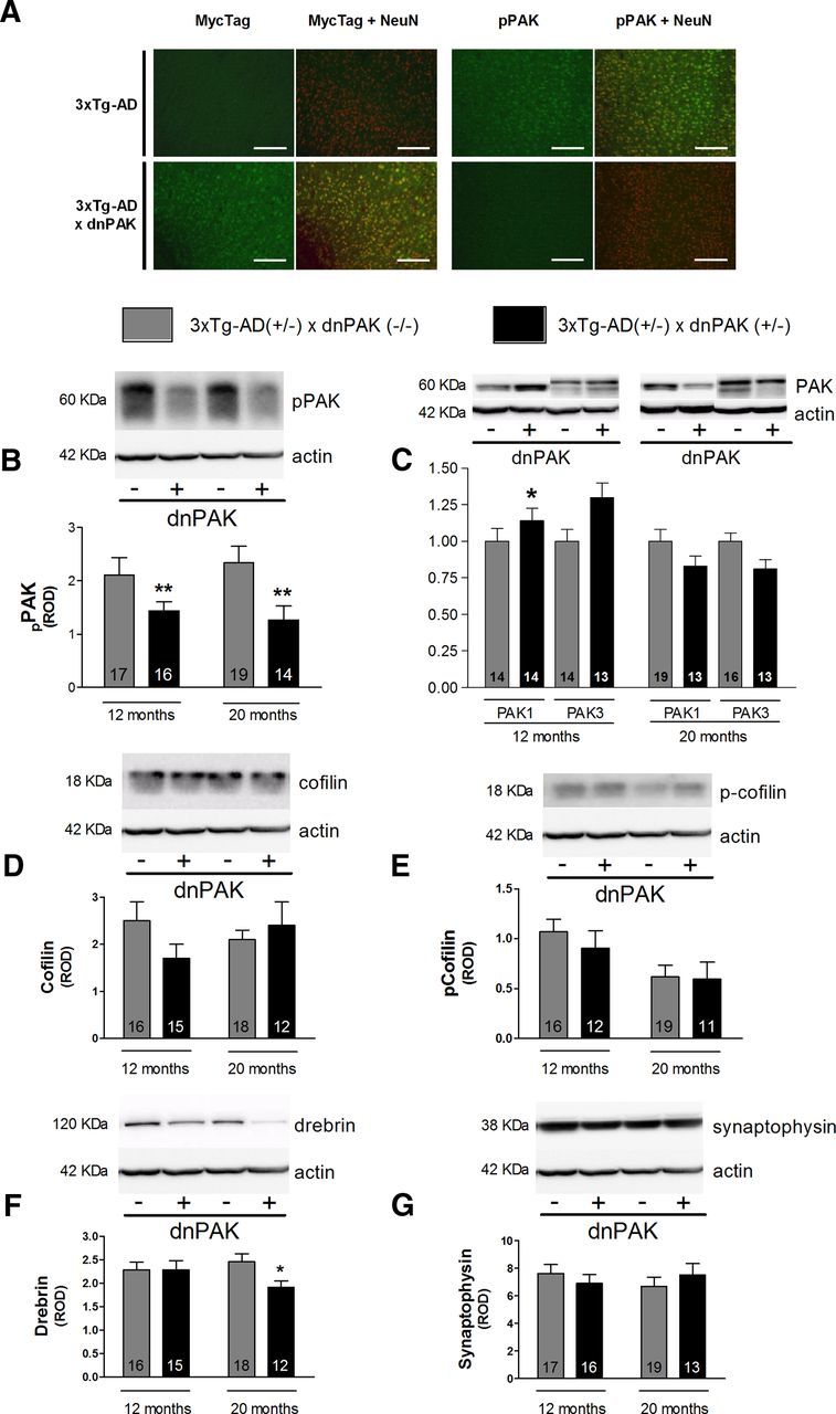
Consequences of PAK inactivation in 12- or 20-month-old heterozygous 3xTg-AD mice. A, Myc-Tag immunofluoresence (green) in the cortex confirmed that the dnPAK transgene was only expressed in neurons (NeuN in red) from 3xTg-AD × dnPAK mice compared with 3xTg-AD mice. pPAK immunostaining (green) in the cortex highlighted a neuronal localization (NeuN in red) of pPAK (s141) and an apparent decrease in 20-month-old 3xTg-AD mice with genetically PAK inactivation compared with 3xTg-AD mice (n = 3 per group). Western blot analyses revealed that dnPAK transgene expression reduced pPAK (B) and increased the level of PAK1 in 12-month-old mice, but was inefficient to modulate PAK3 at both ages (C) in the soluble fraction. dnPAK transgene expression did not induce any effect on cofilin (D) or phospho-cofilin (E) levels in the cortex of 3xTg-AD mice. Interestingly, drebrin level was reduced in 20-month-old mice in the membrane fraction (F). Finally, no significant effect was observed on detergent-soluble synaptophysin level whatever the age of mice (G). Square bar equals 100 μm. Values are expressed as mean ± SEM. Statistical analyses were performed using an unpaired t test. For PAK3 analysis (C), only the upper band was used. *p < 0.05, **p < 0.01.