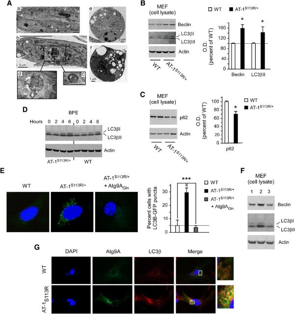
Figure 11.
AT-1S113R/+ mice display abnormal activation of autophagy-MEF studies. A, EM of MEFs. a, MEFs from WT animals. b–f, MEFs from AT-1S113R/+ mice. Different features that are consistent with abnormal activation of autophagy are evident in AT-1S113R/+ MEF cells. A large autolysosome is evident in d, whereas cells undergoing autophagic (type II) cell death are shown in e and f. B, Immunoblots showing increased levels of autophagy markers beclin-1 and LC3β. Error bars indicate SD of n = 3 MEF lines. *p < 0.05. C, Immunoblots showing decreased levels of the autophagy “cargo” protein p62. Error bars indicate SD of n = 3 MEF lines. *p < 0.05. D, Immunoblots showing the autophagy flux in WT and AT-1S113R/+ MEFs. BPE, bafilomycin (500 nm), pepstatin A (10 μg/ml), and E64 (10 μg/ml). E, LC3β-GFP activation and redistribution in AT-1S113R/+ MEF cells. The increased induction of autophagy observed in AT-1S113R/+ MEFs is rescued by expressing the dominant Atg9AGln mutant. Error bars indicate SD of n = 3 MEF lines. ***p < 0.0005. F, Immunoblots showing rescue of the autophagy phenotype after expression of Atg9AGln mutant. Lane 1, WT; lane 2, AT-1S113R/+; and lane 3, AT-1S113R/+ + Atg9AGln. G, Colocalization of Atg9A and LC3β in AT-1S113R/+ MEFs.

