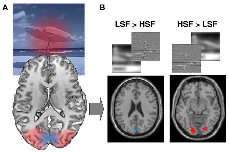
FIGURE 4.
(A) Retinotopic mapping of the visual field on the visual cortex. The central (foveal) part of the visual field is represented at the very back of the visual cortex and laterally. More peripheral regions of the visual field are represented further forward in the medial part of the visual cortex. (B) Retinotopic organization of spatial frequency processing during scene perception: LSF [as opposed to HSF, (LSF > HSF) contrast] scene categorization recruits areas dedicated to peripheral vision, while HSF [as opposed to LSF, (LSF > HSF) contrast] scene categorization recruits areas dedicated to foveal vision. Figure adapted from Musel et al. (2013).

