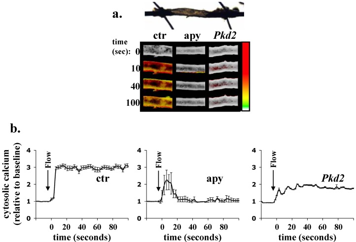
Figure 4.
Calcium response to fluid flow in ex vivo arteries. (a) Within ex vivo arteries, fluid flow causes an influx of calcium. (b) In tissues with knocked-down Pkd2, a ciliary calcium channel, or that are treated with apyrase, the response profile is changed. Tissues were incubated with a fluorescent calcium dye prior to perfusion. Change in cytosolic calcium was pseudocolored; white/green represents a low level of cytosolic calcium, and yellow/red denotes a higher level. Taken and adapted from [6].

