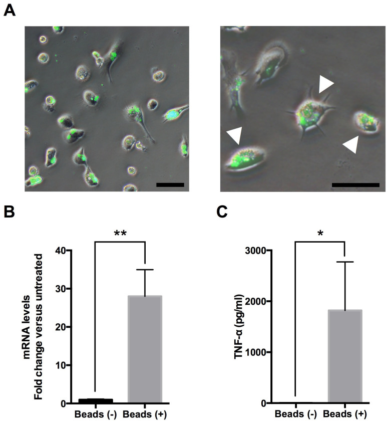
Figure 3. Dynamic functional analysis of the iMG cells.
(A) The iMG cells were incubated with FITC-conjugated latex beads for 24 hours, and phagocytic activity was observed by fluorescent microscopy. The iMG cells showed the ability of phagocytosis with morphological changes into an ameboid form (arrow head). Scale bar, 50 μm. (B and C) The ability of TNF-α production during phagocytosis was measured on the iMG cells. The iMG cells were incubated with latex beads for 72 hours. The extracted RNA and culture supernatant were analyzed by qRT-PCR and Cytometric Beads Array System (CBA), respectively. The mRNA expression (B) and protein level of TNF-α (C) on the iMG cells were significantly higher compared to controls (B, n = 4; C, n = 6). *P < 0.05, **P < 0.01. Error bars, SEM.

