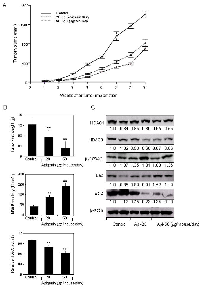
Figure 6.

Effect of apigenin intake on PC-3 tumor growth in athymic nude mice and its correlation with downregulation of HDAC activity and expression. Approximately 1×10⁶ cells were injected into both flanks of each mouse to initiate ectopic prostate tumor growth, and apigenin was provided to the animals 2 weeks before cell inoculation mimicking chemoprevention regimen. Mice were fed ad libitum with Teklad 8760 autoclaved high-protein diet. Apigenin was provided with 0.5% methyl cellulose and 0.025% Tween 20 as vehicle to these animals perorally on a daily basis. Group I, control, received 0.2 ml vehicle only, Group II received 20μg apigenin per mouse in 0.2 ml vehicle and Group III received 50μg apigenin per mouse in 0.2 ml vehicle daily for 8 weeks. Once the tumor xenografts started growing, their sizes were measured weekly in two dimensions throughout the study. A, Tumor volume (cubic millimeter) in control and treated groups B, Wet weight of tumors is represented as the mean of 6–8 tumors from each group, quantitative measurement of apoptosis as demonstrated by M30 reactivity, and relative HDAC activity in PC-3 tumors after apigenin intake at the indicated doses. Values are means ± SD, n = 6–8, repeated twice with similar results. **P < 0.001 versus control. C, Immunoblots for HDAC1, HDAC3, p21/waf1, bax and bcl2 in tumor lysates after apigenin intake at the indicated doses. The blots were stripped and reprobed with anti-β-actin antibody to ensure equal protein loading. The details are provided in ‘materials and methods’.
