
Figure 6.
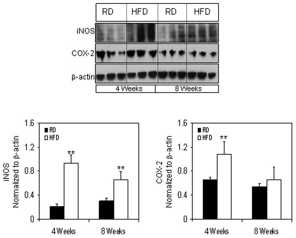
Protein expression of iNOS and COX-2 levels in the total cell lysate extracted from the prostrate of RD and HFD fed NF-κB-Luc mice. (A) Total cell lysates were prepared as described in the ‘materials and methods’ from the prostrate of mice fed with RD and HFD. The samples were subjected to SDS-PAGE gel electrophoresis. The blots were analyzed for the indicated antibodies and β-actin was used as the loading control. Densitometric quantification represents the changes in the levels of iNOS and COX-2 after HFD. Black bars indicate RD and white bars indicate HFD. Data are a mean of ± SD and corrected for loading. The asterisk (**) indicates the significant changes (p<0.01) in HFD compared to its respective RD fed controls.