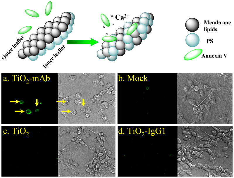
Figure 4.
Laser confocal microscopy images of the localization of Annexin V on outer membrane of the A172 GB cells after treatment with TiO2-mAb and subsequent light exposure. After the light illumination cells were incubated for 6 hours as described in the Supporting Information, permeabilized with Triton X-100 and treated with anti-human FITC-labeled Annexin V. (a) No Annexin V distribution was observed in control experiments: (b) cells with no nanoparticles, (c) cells with bare TiO2 particles, (d) cells with isotype-matched immunoglobulin conjugate TiO2-IgG1.

