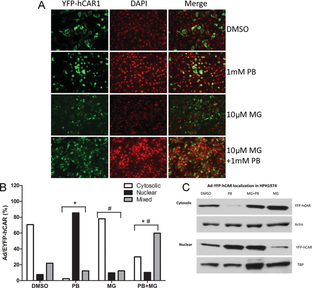
Figure 2. Attenuation of PB-induced nuclear translocation of adenoviral-YFP-hCAR in HPH by MG-132.
HPHs were infected with adenoviral-YFP-hCAR vectors as described in the Experimental section, and treated with vehicle control (0.1 % DMSO), 10 µM MG-132 (MG), and 1 mM PB in the presence and absence of 10 µM MG-132. (A) After 24 h treatment, hepatocytes were DAPI-stained and subjected to fluorescence microscopy analysis. (B) For each treatment, approximately 100 hCAR-expressing cells were counted and classified on the basis of cytosolic, nuclear or mixed (cytosolic plus nuclear) hCAR cellular localization. The percentages of CAR localization on the basis of the microscopy analysis are indicated. Significantly different from DMSO (*), PB (#), P < 0.05. (C) In a separate experiment, cytoplasmic and nuclear fractions were extracted from the treated HPHs for immunoblotting analysis.

