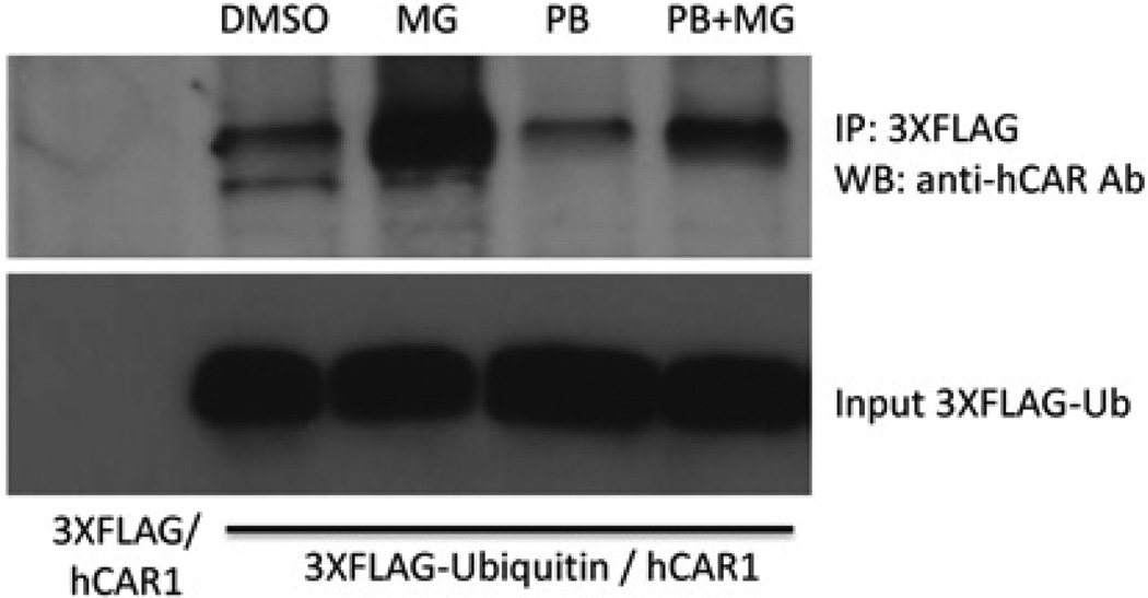
Figure 8. MG-132 increases the ubiquitination of hCAR.
COS-1 cells transfected with hCAR1 expression vector and 3 × FLAG—ubiquitin or 3 × FLAG-empty vector were exposed to chemical treatments for 5 h. Cellular extracts were precipitated with anti-FLAG M2 antibody resin. The resulting total precipitated ubiquitinated protein was subjected to Western blot detection with an anti-hCAR antibody. Lane 1, precipitates from cells transfected with hCAR1 and 3 × FLAG-empty vector; lanes 2–5, precipitates from cells transfected with hCAR1 and 3 × FLAG—ubiquitin after various chemical treatments. The lower panel shows the level of 3 × FLAG—ubiquitin input. Ab, antibody; IP, immunoprecipitation; WB, Western blot; Ub, ubiquitin.

