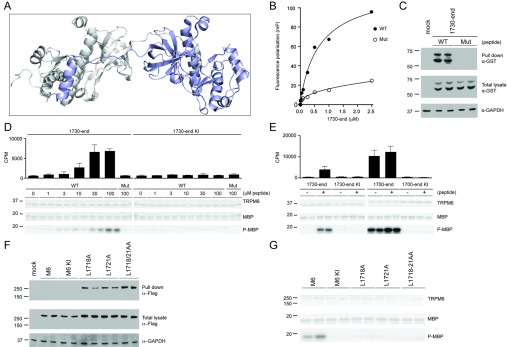
Figure 2. Identification of dimerization motif residues in TRPM6.

(A) Structural model of TRPM7 α-kinase depicting the N-terminal non-catalytic α-helix of one monomer that interacts with the kinase segment of the other monomer. (B) Fluorescence polarization analysis of wild-type (WT) and mutant (Mut) dimerization motif peptides with E. coli-purified GST-tagged TRPM6-(1730–end). (C) GST-tagged TRPM6-(1730–end) was expressed in HEK-293 cells, and lysates were subjected to a peptide pull-down, as described in the Materials and methods section, using either wild-type (WT) or mutant (Mut) dimerization motif peptide. The samples were analysed by immunoblotting with anti-GST antibody (top panel). Total cell extracts were immunoblotted with GST antibody (middle panel), using GAPDH expression as loading control (bottom panel). (D) Immunoprecipitation of FLAG-tagged TRPM6-(1730–end) wild-type and kinase-inactive TRPM6 from HEK-293 cell lysate was followed by a kinase assay using increasing concentrations of the wild-type (WT) dimerization motif peptide, and highest concentrations (100 μM) for mutant (Mut) peptide. Proteins were separated by SDS/PAGE, stained with Coomassie Blue (top and middle gels) and MBP phosphorylation was detected by autoradiography (bottom gel). Incorporated radioactive counts are depicted as a histogram. (E) FLAG-tagged TRPM6-(1730–end) and TRPM6-(1700–end), both wild-type and kinase-inactive (KI) were immunoprecipitated and subjected to a kinase assay without (−) or with (+) 30 μM dimerization motif peptide. Proteins were separated by SDS/PAGE, stained with Coomassie Blue (top and middle gels) and MBP phosphorylation was detected by autoradiography (bottom gel). Incorporated radioactive counts are depicted as a histogram. (F) Full-length FLAG-tagged TRPM6 wild-type, kinase-inactive (KI) and indicated mutants from HEK-293 cells lysate were subjected to a peptide pull-down, as described in the Materials and methods section, using the wild-type dimerization motif peptide. The pull-down samples were analysed by immunoblotting with anti-FLAG antibody (top panel). Total cell extracts were immunoblotted with anti-FLAG antibody (middle panel), using GAPDH expression as loading control (bottom panel). (G) Immunoprecipitated FLAG-tagged TRPM6 and indicated mutants were subjected to a kinase assay using MBP as substrate. Proteins were separated by SDS/PAGE, stained with Coomassie Blue (top and middle panels) and MBP phosphorylation was detected by autoradiography (bottom panel). Molecular masses are indicated in kDa.
