Fig. 1.
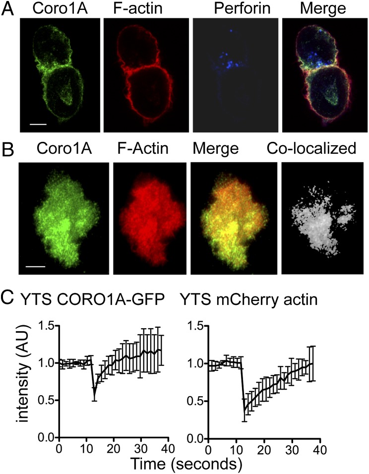
Coro1A localizes to the NK cell cytolytic IS. (A) YTS NK cells conjugated to 721.221 target cells were fixed, permeabilized, and stained for Coro1A (green, Far Left), F-actin with phalloidin (red, Left Center), and perforin (blue, Right Center). (B) YTS-CORO1A-GFP mCherry-actin cells activated by immobilized antibody on glass were imaged at one frame per minute by TIRFm. Shown are representative images 10 min after contact with the imaging chamber. (Far Right) Colocalized pixels were calculated and are shown in grayscale. (C) YTS-CORO1A-GFP mCherry-actin cells were activated with immobilized antibody for 10 min, and fluorescence was bleached to 50% for both GFP and mCherry fluorophores. Fluorescent intensity for both channels was normalized, and the mean ± SD is shown for eight cells from three independent experiments. AU, arbitrary units. (Scale bars: 5 μm.)

