Fig. 6.
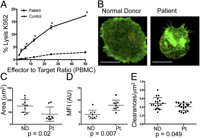
NK cells from a Coro1A-deficient patient have defective cytotoxic function and altered F-actin architecture. (A) Peripheral blood mononuclear cells (PBMCs) from the patient (dashed line) or a healthy donor control (solid) were incubated with susceptible K562 targets, and specific lysis of targets was measured by standard 51Cr-release assay. *P < 0.05 by Student's two-tailed unpaired t test. (B) NK cells from a normal donor (Left) or the patient (Right) were enriched by negative selection and analyzed by STED nanoscopy at the plane of the glass as in Fig. 3. Shown is one representative cell from two independent experiments. (Scale bars: 5 μm.) (Resolution: 110 nm.) Area (C, square micrometers) and MFI (D) of F-actin were measured as in Fig. 3 (n = 10 from three independent experiments). (E) F-actin clearances per square micrometer (250–500 nm) were measured for cells from the normal donor (●) and the patient (■). Each data point represents clearances per square micrometer for one cell (n = 18 cells per condition). ND, normal donor; Pt., patient.

