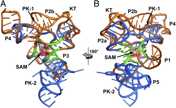
Fig. 3.
Superimposition of the TteSAM-I and env87SAM-I/IV aptamer domains. (A) The TteSAM-I riboswitch structure (Protein Data Bank accession code 2GIS) is shown in orange, and the env87SAM-I/IV structure in blue. The conserved core between the two RNAs is emphasized in green. This core represents the bases used to align the structures. (B) 180° clockwise rotation of the aligned structures.

