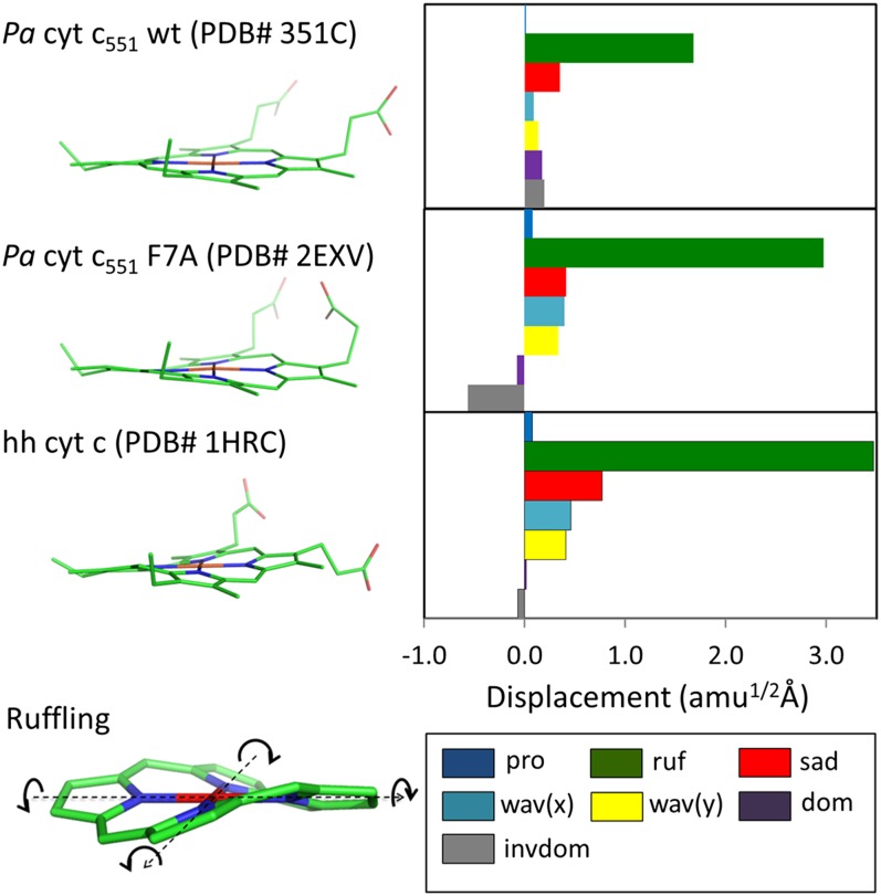
Fig. 1.
Crystal structure and NSD analysis of hemes in ferric Pa cyt c551 and its F7A mutant are compared with hh cyt c. The minus sign of displacement is defined only for doming and inverse doming to indicate the direction of Fe displacement (+, proximal; −, distal). The ruffling mode is shown at the lower left part of the figure and the arrows indicate the rotation of pyrrole rings with respect to Fe–N axis (dotted black lines).

