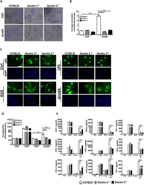
Figure 2. The role of Dectin-1 and Dectin-2 in macrophage activation by ΔrodA and HF treated conidia.
A. C57Bl/6, Dectin-1−/−, and Dectin-2−/− bone marrow macrophages were plated on coverslips and incubated 1h with ΔrodA or G10 conidia. Original magnification is x100, and the size bar is 10 μm. B. Mean+/-SD associated conidia per macrophage C. p65 translocation to the nucleus of C57Bl/6, Dectin-1−/−, and Dectin-2−/− bone marrow derived macrophages after 1h incubation with ΔrodA conidia. Macrophages were fixed, permeabilized, and stained with anti-p65 primary antibody and Alexafluor-488 tagged anti-rabbit secondary antibody. Cells were visualized by fluorescent microscopy using oil immersion (size bar is 10 μm). D. Nuclear translocation was quantified by image analysis using Metamorph software. E. CXCL1, CXCL2 and TNF-α production by C57Bl/6, Dectin-1−/−, and Dectin-2−/− bone marrow derived macrophages after 6h incubation with ΔrodA or HF treated conidia at a ratio of 1:50 (MOI=50). Cytokine production was measured by ELISA. P values are: *<0.05, **<0.001, ***<0.0001. Experiments were performed at least twice with similar results.

