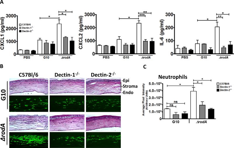
Figure 5. The role of Dectin-1 and Dectin-2 in the early host response following corneal infection with ΔrodA and G10 conidia.
A. Cytokine production in the cornea at 6h post-infection. C57Bl/6, Dectin-1−/−, and Dectin-2−/− mice were injected intrastromally with 5×10⁴ G10 or ΔrodA conidia as before, and after 6h, corneas were dissected and homogenized, and CXCL1/KC, MIP-2, and IL-6 were measured by ELISA. B,C. Histological sections (5 μm) were stained with PASH, or immunostained with NIMP-R14 to detect neutrophils. B. Representative sections near the peripheral cornea at the site of neutrophil infiltration. Image bar represents 40X magnification, size bar is 20 μm. C. Quantification of NIMPR-14 staining by image analysis (Metamorph) shows average pixel intensity of the entire corneal section (mean+/-SD of five mice per group). P value is *<0.05, **<0.001, ***<0.0001. Experiments were repeated twice with similar results.

