Figure 3.

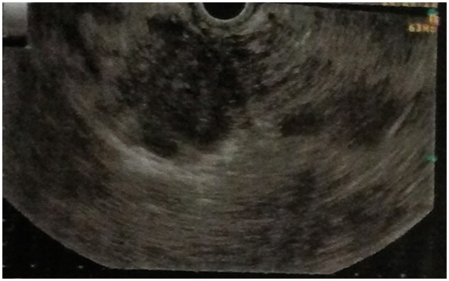
EUS image showing hypoechoic lobulated mass in relation to the pancreatic head, with anechoic areas and floating echogenic material within.

EUS image showing hypoechoic lobulated mass in relation to the pancreatic head, with anechoic areas and floating echogenic material within.