
Figure 4.
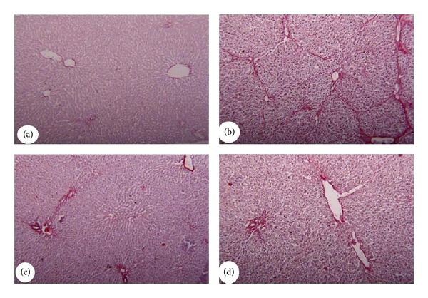
Photomicrographs of liver tissue stained with Sirius Red showing. (a) Control group showing absence of collagen fibers. (b) CCl4 group had developed extensive fibrosis in the periportal area and more fibrillar collagen deposition. (c) Physalis group showing absence of collagen fibers. (d) Physalis + CCl4 group showing sporadic, small fibrotic lesions in the periportal zone and reduction in collagen fibers deposition. Original magnification 200x.