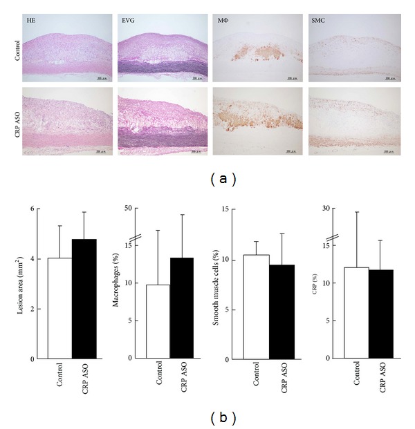
Figure 5.

Microscopic analysis of the aortic lesions. Representative micrographs of the aortic lesions from CRP ASO-treated and control rabbits (a). Serial paraffin sections were stained with hematoxylin-eosin (HE) and elastica van Gieson (EVG) or immunohistochemically stained with monoclonal antibodies (mAbs) against either macrophages (Mφ) or α-smooth muscle actin for smooth muscle cells (SMC) or rabbit CRP. Intimal lesions on EVG-stained sections and positively immunostained areas of macrophages; SMC and CRP were quantified with an image analysis system (b). n = 9 for each group.
