
Figure 3.
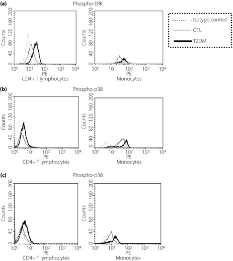
Representative histograms of flow cytometry analysis of phospho‐mitogen‐activated protein kinases including (a) phospho‐extracellular signal‐regulated kinase (ERK), (b) phospho‐p38, and (c) phospho‐c‐Jun NH2‐terminal protein kinase (JNK) in CD4+ T lymphocytes and monocytes from peripheral blood mononuclear cells in patients with type 2 diabetes (T2DM) and the control group (CTL). Triplicate experiments were carried out with essentially identical results and representative figures are shown. Immunoglobulin G1 isotypic control antibodies, which have no specificity for target cells within a particular experiment yet retain all the non‐specific characteristics of the antibodies used in the experiment. Inclusion of this antibody is to confirm the specificity of primary antibody binding and exclude non‐specific fragment crystallizable receptor binding to cells or other cellular protein interactions.