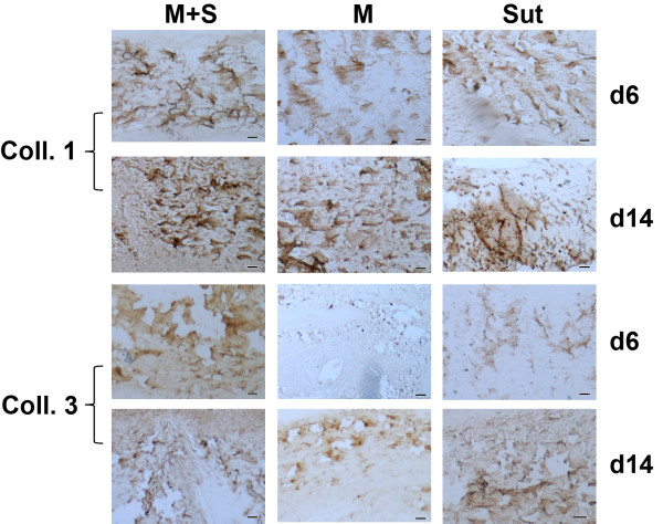
Figure 4.
Immunohistochemistry of types I and III collagen. In general, type I collagen was stained in larger fibers, while type III collagen was detected on finer fibers in the repaired tendons. The distribution of types I and III collagen was very similar among the groups, except of noticeable weak staining of type III collagen in the M group at day 6 (bar = 50 μm).

