
Figure 1.
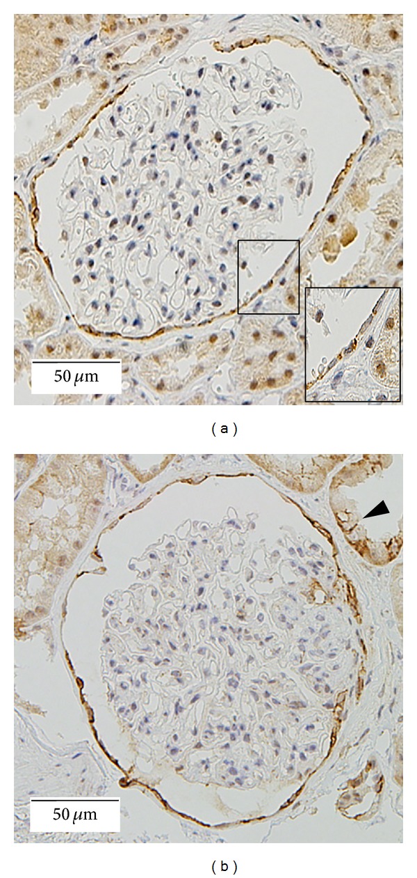
Claudin-1 stainings for nephrectomy ((a) ×200) and minimal change nephrotic syndrome ((b) ×200) samples are shown. In glomeruli, claudin-1 shows a strong, sharp staining pattern along cell membranes of adjacent PECs (magnified view in (a)). Staining signals are mainly detected at cell to cell contact sites. Distal tubules also express claudin-1 ((b) arrow head).