
Figure Figure 4.
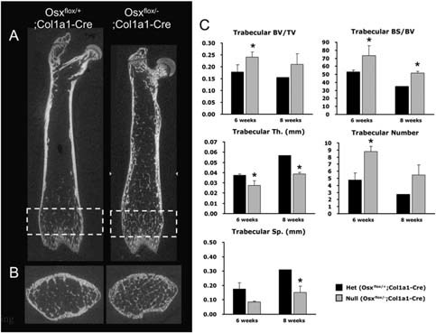
Analysis of trabecular bone architecture in Osx flox/–;Col1a1‐Cre. (A) 2D longitudinal image by μCT analysis of femoral bone at 6 wk of age. Osteopenic cortical bone and small pieces of trabecular bones were observed in Osx flox/–;Col1a1‐Cre. (B) μCT analysis of rectangled metaphysis in A. Increased immature or premature trabecular bones were observed with the osteopenic cortical bone in Osx flox/–;Col1a1‐Cre. (C) Histomorphometrical analysis of trabecular bone at 6 and 8 wk of age. Compared with Osx flox/+;Col1a1‐Cre mice, trabecular bone volume (BV/TV) and bone surface (BS/BV) were increased in Osx flox/–;Col1a1‐Cre mice, accompanied by increased trabecular numbers. However, trabecular thickness (Th) and separation (Sp) were significantly reduced in Osx flox/–;Col1a1‐Cre, indicating immature or premature trabecular bones. *p < 0.05.