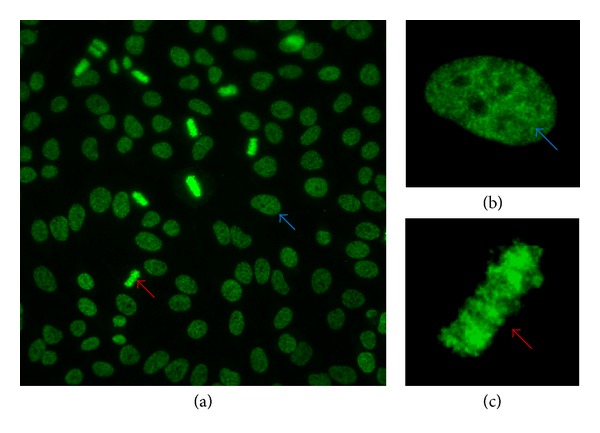
Figure 3.

Characteristic staining pattern of anti-DFS70 antibodies. The characteristic dense fine speckled (DFS) staining pattern of interphase HEp-2 cells is indicated by the blue arrow and the strong chromatin staining of mitotic cells by the red arrow. (a) Wide field view using 40x magnification, (b) dense fine speckled pattern of an interphase nucleus, and (c) of the metaphase chromatin of a mitotic cell.
