Figure 7.
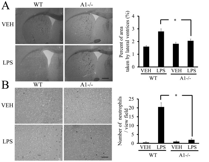
Plexin-A1−/− mice show reduced ventricular enlargement and neutrophil infiltration after LPS injection. (A) LPS injection leads to the enlargement of lateral ventricle in WT mice, but not in Plexin-A1−/− mice. Scale bar is 1,000 μm. Quantification of the area of the lateral ventricle revealed a significant increase in WT mice treated with LPS as compared with saline-treated mice. By contrast, there was no significant increase in lateral ventricular area in the LPS-treated Plexin-A1−/− mice as compared with saline-treated Plexin-A1−/− mice. (B) Esterase staining to detect neutrophil was performed with brain sections from mice injected with saline or LPS. Saline administration to WT mice did not induce any infiltration of neutrophil detected by esterase staining in the cerebral cortex, while WT mice injected with LPS show many infiltrating neutrophils in the cortical area. Esterase staining hardly detects neutrophil in the cerebral cortex even after administration of LPS to Plexin-A1−/− mice. Quantification of neutrophil number shows a significant increase in neutrophil in the cerebral cortex in LPS-treated WT mice as compared with saline-treated WT mice. By contrast, there was no significant increase of neutrophil infiltration into the cerebral cortex in LPS-treated Plexin-A1−/− mice compared with saline-treated control mice. Results are shown as means ± SEM, *p<0.05. VEH, vehicle (saline); LPS, lipopolysaccharide; WT, wild-type; A1−/−, Plexin-A1−/−.

