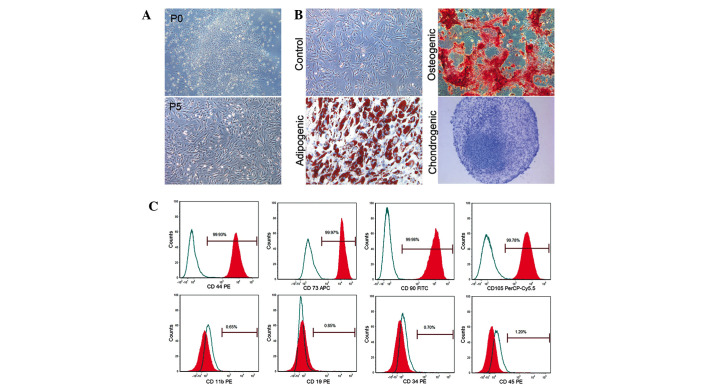
Figure 1.
Characterization of rat BMSCs. (A) Spindle-shaped cells formed a colony in primary cells and cells after passaging several times (P5) remained with their fibroblast-like morphology (magnification, ×40); (B) Differentiation potentials of BMSCs: osteogenic differentiation (Oil Red O staining; magnification, ×100), adipogenic differentiation (Alizarin Red S staining; magnification, ×200) and chondrogenic differentiation (Toluidine Blue staining; magnification, ×100). (C) Several markers of BMSCs identified by flow cytometry. BMSCs, bone marrow mesenchymal stem cells.

