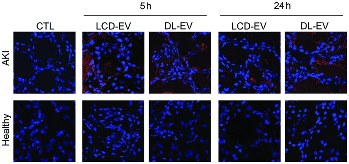
Figure 7.
Confocal microscopy of fluorescent cell-derived extracellular vesicles (EVs) in kidneys. Representative micrographs of kidney sections of mice with acute kidney injury (AKI) and healthy mice sacrificed at 5 and 24 h after EV injection (red). Nuclei were counterstained with Hoechst dye. Two kidney specimens were analyzed for each experimental point. Original magnification, ×630. LCD-EV, labeled EVs produced by donor cells; DL-EV, directly labeled EVs.

