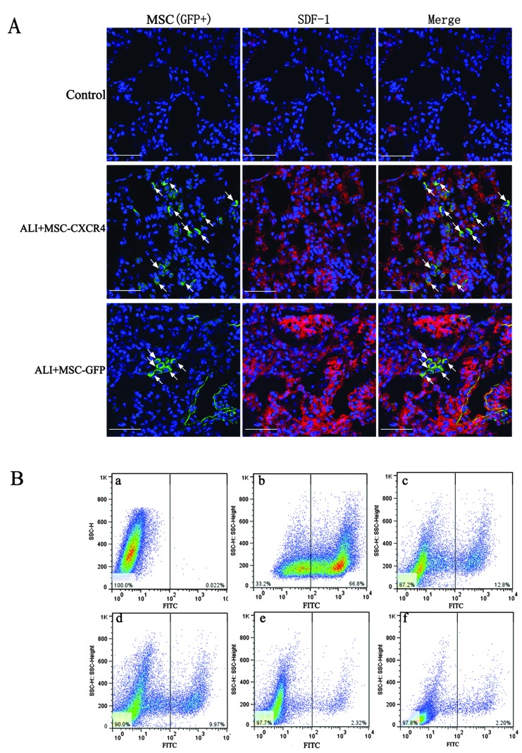
Figure 3.
Immunofluorescent staining and flow cytometric analysis was used to detect the engraftment of mesenchymal stem cells (MSCs) (white arrows, green fluorescent cells, GFP+ cells) in lung tissue. (A) Immunofluorescent staining detected the engraftment of MSCs. No GFP+ cells (MSCs) are detectable in the control lung tissue, while the expression of stromal cell-derived factor (SDF)-1 (red) is very low. GFP+ cells are detectable in acute lung injury (ALI)+MSC-CXC chemokine receptor (CXCR)4 and ALI+MSC-GFP groups. More GFP+ cells were identified at SDF-1-expressed sites in the ALI+MSC-CXCR4 group compared with the ALI+MSC-GFP group. Scale bar, 50 μm. (B) Flow cytometric analysis for the engraftment ratio of MSCs in lung tissue. MSCs transfected with Ad-CXCR4 or GFP reporter gene (Ad-GFP) were termed GFP+ cells. Flow cytometry was used to analyze the GFP+ cells in lung cell populations subsequent to MSC administration into lung, as shown in the right area. The SDF-1/CXCR4 axis promotes MSCs to engraft into injured lung tissue. (a) Control group with GFP+ cell injection; (b) MSCs transfected with Ad-GFP is used as positive control; (c) MSC-CXCR4 cells injection in ALI lung on day 7 post-transplantation; (d) MSC-GFP cells injection in ALI lung on day 7 post-transplantation; (e) MSC-CXCR4 cells injection in ALI lung on day 14 post-transplantation; (f) MSC-GFP cells injection in ALI lung on day 14 post-transplantation. More GFP+ cells were detected in the ALI+MSC-CXCR4 group compared with the ALI+MSC-GFP and control groups.

