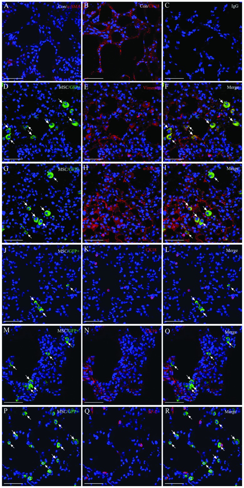
Figure 6.
Detection of mesenchymal stem cell (MSC) differentiation 14 days after transplantation in injured lung. Engraftment of MSCs in lung shown as GFP+ (green fluorescent cells, white arrows) and antibodies to specific cell-type markers (red); co-localization in each case appears yellow. (A–C) Normal control group for α-SMA, CK18 and IgG. (D–R) Immunofluorescent staining for the engraftment of MSC differentiation in the ALI+MSC-CXC chemokine receptor (CXCR)4 group demonstrated that MSCs expressed myofibroblast or fibroblast markers, but did not express epithelial markers. Engraftment of MSCs was almost differentiated into myofibroblasts or fibroblasts, however, rarely differentiated into lung epithelial cells. Nuclear staining was performed using 4′,6-diamidino-2-phenylindole (DAPI). Scale bar, 50 μm.

