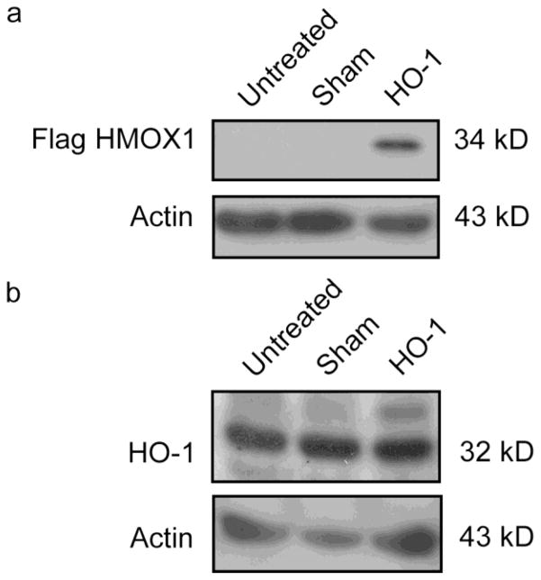
Fig. 1. Expression of Flag-tagged HO-1 at 3 days post-transfection.
(a) Western blot of Flag-HO-1 for untreated, sham transfected, and HMOX1-transfected cells at 3 days post transfection. Flag-tagged HO-1 is detectable in HMOX1-transfected cells but not in control preparations. (b) Western blot of endogenous HO-1 for untreated, sham-transfected and HMOX1-transfected cells at 3 days post transfection. Flag-tagged HO-1 is only detectable in HMOX1-transfected cells as a 34 kDa band approximately 2 kDa heavier than endogenous HO-1. The transgene yields product that is immunopositive for both the Flag peptide and HO-1 protein

