
Figure 4.
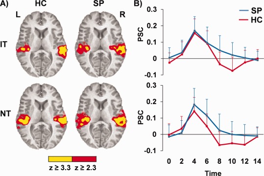
Panel (A) shows auditory activation for IT and NT tones relative to baseline for a. controls (HC) and patients (SP) (blue) and are color‐coded according to magnitude of the z scores. Graphs in Panel (B) illustrate aberrant HRFs (postpeak) for b. SP (blue) compared to HC (red) in bilateral auditory cortex for identical (IT; upper graph) and nonidentical (NT; lower graph) tones, with percent signal change (PSC) represented along the y‐axis. Error bars represent 2 × standard error of the mean.