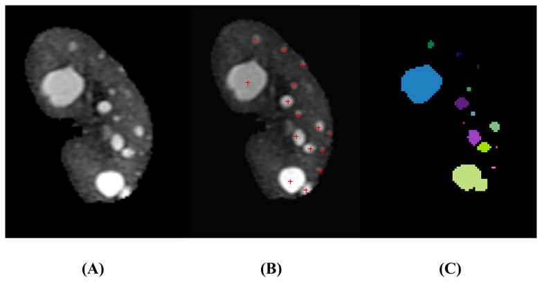
Fig. 1. Mid-slice MR images from an ADPKD patient with mild cyst-burden illustrating (A) the initial segmented kidney, (B) manual cyst counting, and (C) individual cysts segmented using the semi-automated method.

(A) Cysts were scattered and discretely defined by surrounding renal parenchyma. Most cysts were well separated without touching each other. (B) The manual counting method was performed using an in-house cyst-labeling computer program. Small poorly-defined foci of faint brightness were not counted as cysts because they were difficult to differentiate from background MR image noise in the parenchyma. (C) Individual renal cysts were segmented and color-coded using the semi-automated method.
