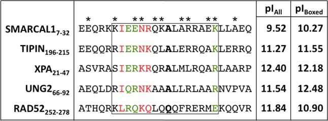
Figure 7.

Sequence alignment of RPA32C target interaction motifs. The asterisks above the SMARCAL1-RMB sequence identify the SMARCAL1-RBM residues in contact with RPA32C in our RosettaDock model. Residues colored green and red represent those that are conserved and highly conserved, respectively. The residues corresponding to the critical alanine residue at position 14 in SMARCAL1 are highlighted in bold. The box is drawn to show the residues that correspond to the RPA32C binding region in the NMR structure of the complex with UNG2. The two columns at right list the pI values of all residues in the motif (pIall) and of only residues in the box (pIbox). The alignment was generated using ClustalW.45
