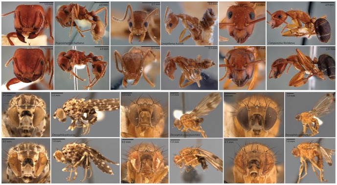
Figure 3. Images of representative specimens before and after extraction.
Specimens before extraction are in first and third rows, and after extraction in second and fourth rows. For ant photos, the same specimen is depicted before and after extraction. For Drosophila, different specimens from the same collection series were used for the before-and-after comparison. Specimen damage to ant was minimal, consisting only of eye de-pigmentation. The more fragile Drosophila specimens were more greatly affected, and their eyes show signs of partial collapse. The more fragile fruit fly specimens also showed signs of greater mechanical damage due to handling.

