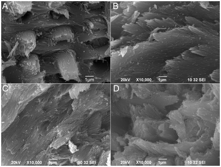
Figure 4. Scanning electron microscope (SEM) images of tooth enamel.
Teeth were cracked open with a diamond knife and then coated with gold/platinum for SEM observation. Panel A is taken from a normal (wild-type) incisor taken from a mature NBCe1+/+ animal. Panels B, C and D are taken from the NBCe1+/+, NBCe1+/− and NBCe1−/− teeth, respectively, grown in the kidney capsule. Scale bars are included in each panel.

