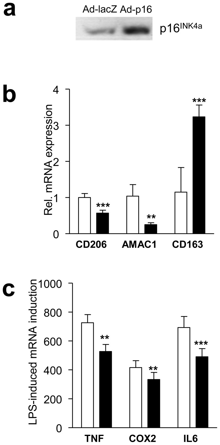
Fig. 3.
Overproduction of p16INK4A in MDMs diminishes M2 polarisation and the LPS-induced inflammatory response. MDMs were infected with either control LacZ or p16INK4A. a p16INK4A protein was detected by western blot. b,c After infection, mRNA was isolated and expression of CD206, AMAC1 and CD163 (b) or LPS-induced TNF, COX2 and IL6 (c) was quantified by quantitative RT-PCR. Statistically significant differences are indicated (t test; ***p<0.001 and **p<0.01). Ad-LacZ, MDMs infected with control lacZ, white bars; Ad-p16, MDMs infected with p16INK4A adenovirus, black bars

