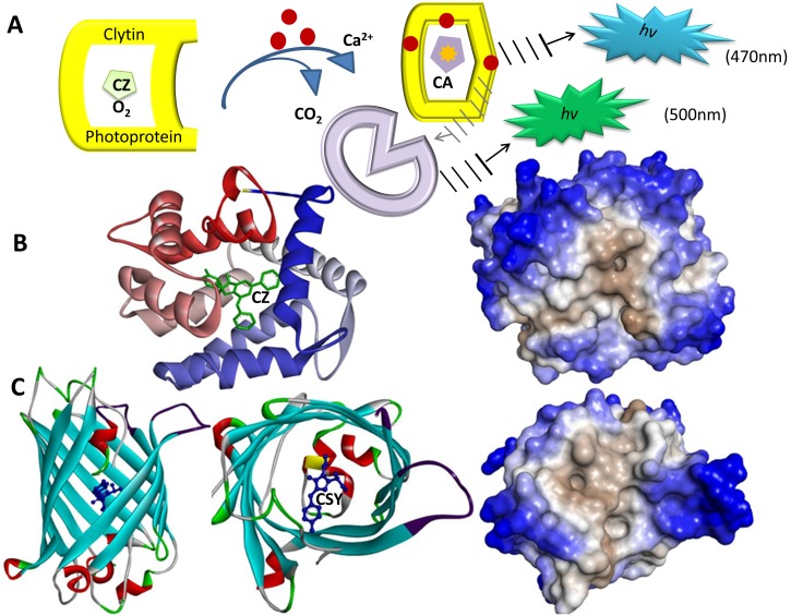
Figure 7. Bioluminescence system of the jellyfish Clytia with the GFP-clytin complex.
(A) Clytin is a Ca2+-regulated photoprotein comprising coelenterazine (CZ) as a substrate for its bioluminescence reaction. When Ca2+ binds to clytin, the bioluminescence reaction is triggered to produce the excited state product coelenteramide (CA) and CO2 with emission of a broad blue bioluminescence (maximum 470 nm). Due to energy transfer, Clytia GFP fluorescence is in green (maximum 500 nm). (B) The structure of clytin (22.4 kDa, PDB code 3KPX) and its representation using hydrophilic (outer part in blue) and hydrophobic (inner part) residues. (C) The structure of Clytia GFP (PDB code 2HPW) and its representation using hydrophilic and hydrophobic residues using Discovery Studio 3.5. The visible fluorophore of Clytia GFP (CSY) is a sequence of three amino acids (68S, 69Y, and 70G). To protect the chromophore fluorescence from quenching by water, the tightly packed nature of the barrel excludes solvent molecules resulting in that the environment comprising this fluorophore is also hydrophobic.

