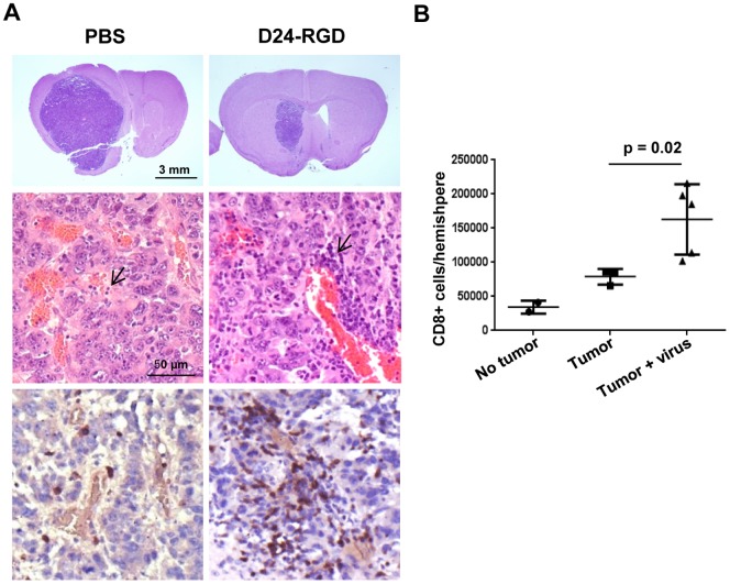
Figure 3. Delta-24-RGD treatment mediated tumor shrinkage and lymphocytic infiltration.
20 days after GL261 implantation and 9 days Delta-24-RGD injections, as indicated in Figure 2, the murine brains were fixed (A) or the leukocytes from fresh hemispheres with or without tumor were isolated and analyzed with flow cytometry (B). A. Brains were stained with hematoxylin-and-eosin (upper two panels) or immune stained for CD3 (bottom panel). Note that, as expected, lymphocytes as round cells with little visible cytoplasm and dense and monochromatic nucleus (arrows) are more prominent in perivascular areas. B. CD8+ T cells were quantified and data represented as mean ± S.E.; n = 3–4. p = 0.02 (Student's t-test, double sided). No tumor: naïve mice; Tumor: GL261-glioma bearing mice; Tumor + virus: mice with Delta-24-RGD-infected GL261-glioma bearing mice. Note that the viral injections resulted in accumulation of lymphocytes, and, importantly, a significantly higher number of CD8+ cells at the tumor site.

