
FIGURE 10.
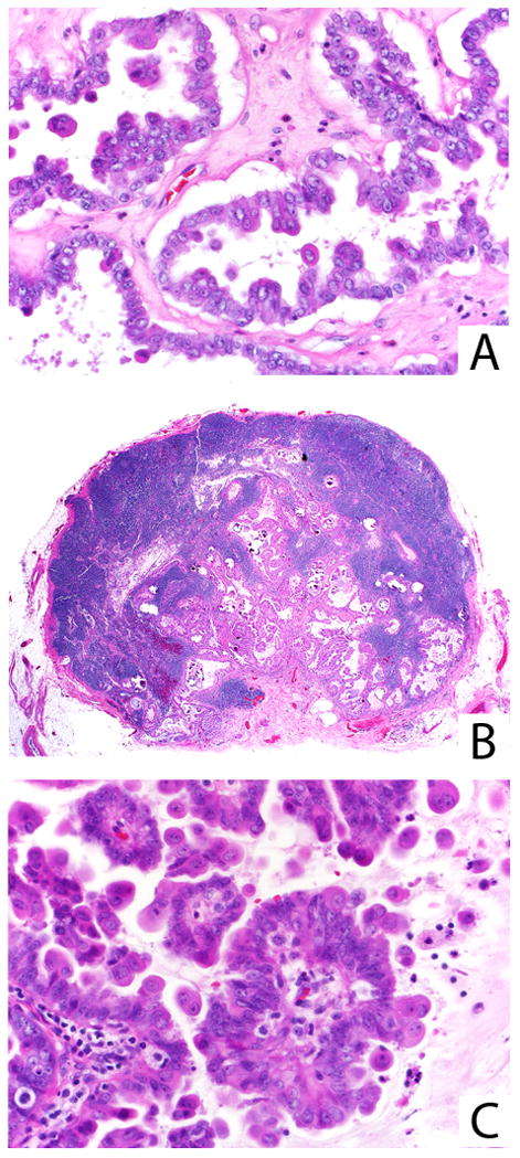
Ovarian SBT/APST with prominent eosinophilic cells (A). The associated lymph node is extensively involved by tumor (B), which on higher power appears identical to the ovarian tumor (C). Both cuboidal/columnar and eosinophilic cells are present in both sites.