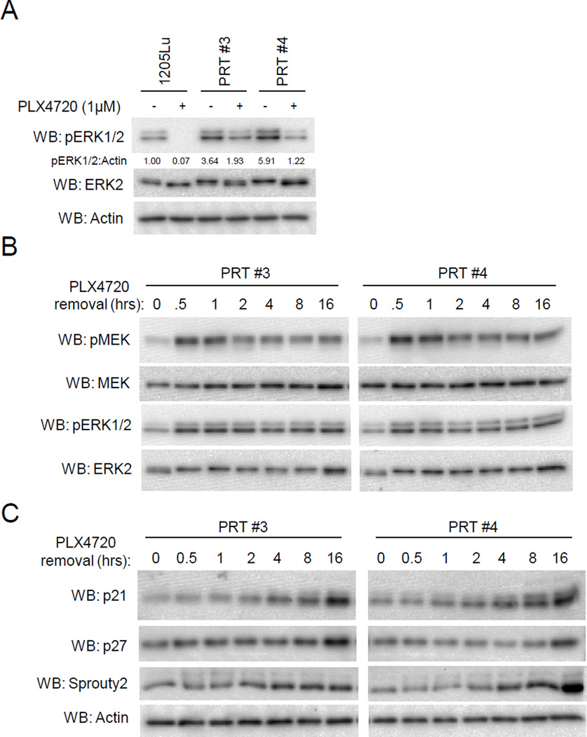
Fig. 3. Phospho-ERK1/2 levels are hyperactivated in PRT #3 and PRT #4 cell lines after RAF inhibitor withdrawal.
(A) Parental 1205Lu cells were seeded overnight in drug-free medium, and PRT #3 and PRT #4 cells were seeded in 1µM PLX4720. The next day, cells were washed and then administered either 1µM PLX4720 or DMSO for a 16 hour treatment. Western blots were performed to assay ERK1/2 activation status. (B) PRT #3 and PRT #4 cells were cultured in PLX4720 (1µM) and then medium changed to drug-free medium. Cell lysates were harvested at the indicated time and analyzed by western blotting for levels of phosphorylated MEK as well as phosphorylated ERK1/2. (C) Similar to (B), cells were lysed after culturing in drug-free medium for the indicated time, and the CDK inhibitors p21Cip1 and p27Kip1 were analyzed by western blot, as well as the putative ERK target sprouty2.

