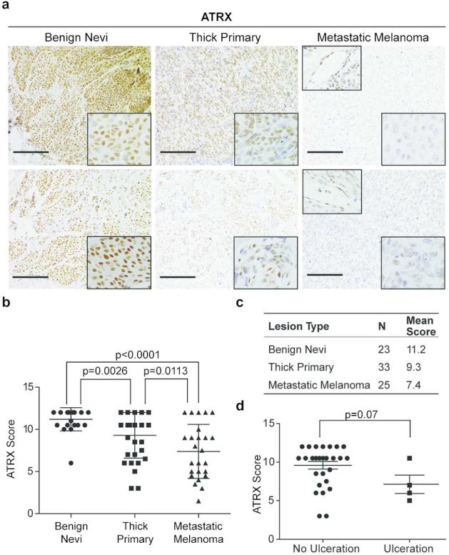
Figure 1. Loss of ATRX protein expression is associated with melanoma progression.
(a) IHC for ATRX in representative benign nevi, thick primary, and metastatic melanoma tissue. Images were taken at 20x magnification; insets (bottom right) show nuclei at 40x magnification. Insets (top left) show ATRX positive stain of endothelial cells within metastatic specimens. Scale bar 100 μm. (b) IHC scores of benign nevi, thick primary, and metastatic melanoma from two independent pathologists. Each tissue section was quantified based on number of positively stained cells (1-4) multiplied by stain intensity (1-3) to generate a score. (c) Table summarizing number of total samples and average IHC score per lesion. (d) ATRX protein levels vs. presence of ulceration in primary melanoma specimens. All statistical significance assessed using two-side Mann-Whitney U test, p value indicated. Mean +/- SD.

