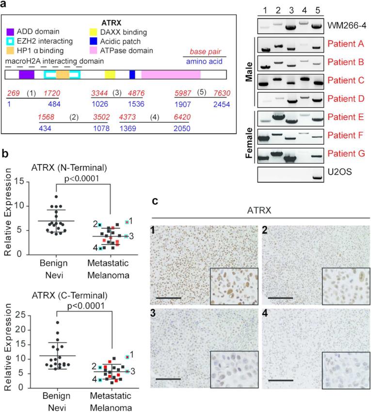
Figure 2. ATRX mRNA levels are decreased in metastatic melanoma.
(a) Illustration of ATRX with domains and five amplicons depicted (left). ATRX cDNA was amplified as indicated for analysis of putative deletions in metastatic melanoma specimens (right). WM266-4 and U2OS were used as controls. (b) qRT-PCR analysis of ATRX from fresh frozen benign nevi and metastatic melanoma lesions. N- and C-terminal primers were used. Melanoma specimens analyzed in (a) are depicted in red and in (c) are highlighted in blue and numbered. Expression levels were normalized to GAPDH and statistical significance was derived using unpaired Student's t-test, p-values indicated. Mean +/- SD. (c) ATRX IHC in representative metastatic melanoma tissues from (b). Images were taken at 20x magnification; insets (bottom right) show nuclei at 40x magnification. Scale bar 100 μm.

