Figure 9.
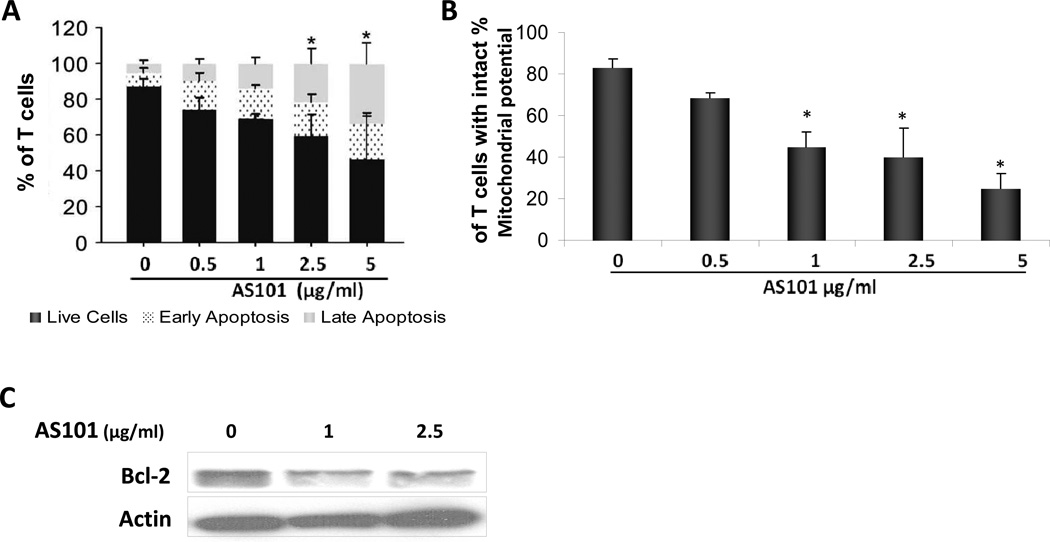
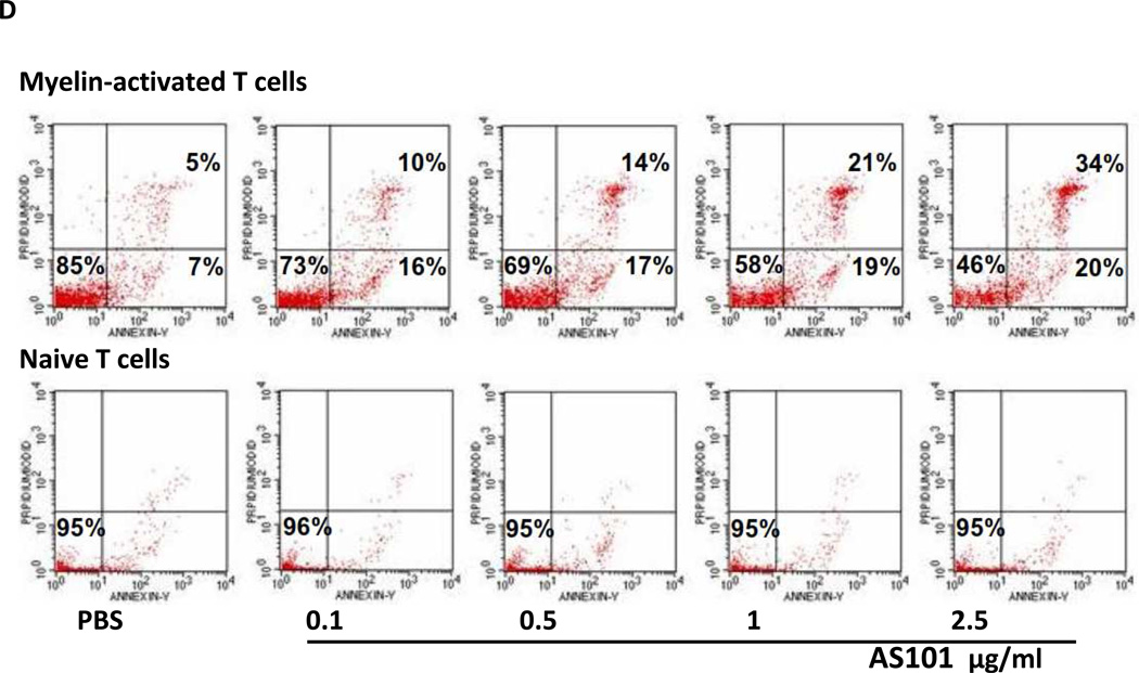
AS101 induces apoptosis in MS patient-derived T-cell lines. (A) Patient-derived T cell lines were incubated with AS101 at the indicated concentrations. After 72 hours, cells were stained with Annexin V and PI, and the percentage of live cells (Annexin V−, PI−), cells in early apoptosis (Annexin V+, PI−) and in late apoptosis (Annexin V+, PI+) were determined by FACS. The means ± SD of three independent experiments are shown. (*P<0.05) (black- live cells, dotted-early apoptosis, gray- late apoptosis). (B) The effect of AS101 on mitochondrial transmembrane potential of myelin antigen-reactive T cells. T cells were treated with AS101 at the indicated concentrations for 72 hr. The cells were incubated with rhodamine 123, and cell fluorescence reflecting the membrane potential, was analyzed by FACScan. (C) Activated T cells were incubated with AS101 (1 µg/ml and 2.5 µg/ml). After 48 hours, the cells were lysed and the levels of Bcl-2 were determined by western blot analysis. (D) Comparison of the effect of AS101 on patient-derived myelin antigen-reactive T-cells vs. naive T cells from normal donors. AS101 was administered at the indicated concentrations, to both activated and naive T cells. The percentage of live cells, cells in early apoptosis, and cells in late apoptosis were analyzed by FACScan. Myelin-activated T cells (upper panel) and naive T cells (lower panel) following treatment with AS101 alone for 72 hours. Results shown are representative from at least three different patients in each group.


