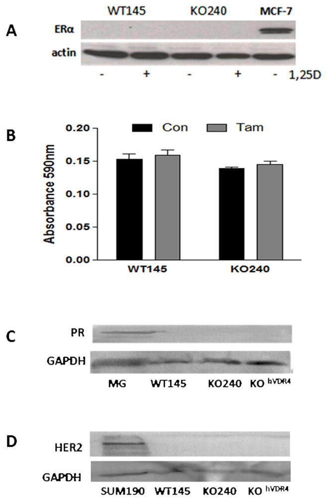
Figure 3. WT145 and KO240 cells are triple negative.

A, Western blot for ERα (top) in lysates from WT145 and KO240 cells. MCF7 breast cancer cell lysate was used as positive control. Blots were stripped and re-probed with actin antibody was used to verify protein loading. B. Effect of tamoxifen on WT145 and KO240 cell density. Cells were treated with ethanol vehicle control (Con) or 100nM tamoxifen (Tam) (bottom) for 96h and analyzed by crystal violet staining for adherent cell density. C. Western blot for progesterone receptor (PR) in WT145, KO240 and KOhVDR cell lysates; homogenate from pregnant mouse mammary gland (MG) was used as positive control. Blots were stripped and re-probed with Gapdh antibody to confirm protein loading. D. Western blot for Her2 in WT145, KO240 and KOhVDR cell lysates; lysate of known HER2+ human breast cancer cell line SUM190 was used as positive control. Blots were stripped and re-probed with Gapdh antibody to confirm protein loading.
